FIGURE 2.
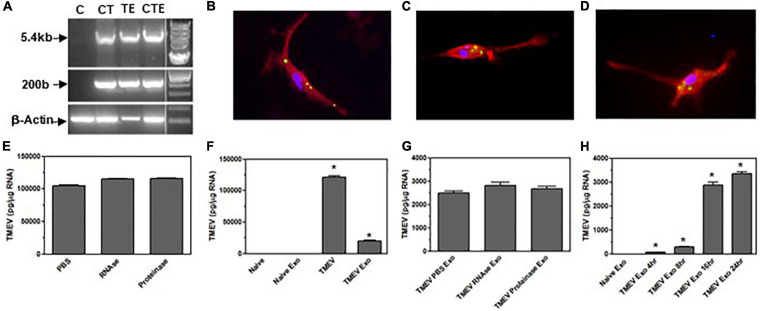
Exosomes from TMEV-infected microglia contain viral RNA that is transferred to bystander microglia. (A) Microglia were infected with TMEV and exosomes were isolated (lane TE). Microglia (1 × 10⁶) were uninfected (lane C), infected with TMEV (lane CT), or incubated with exosomes (100 μg) from TMEV-infected microglia (lane CTE). The cells were lysed 24 h later, RNA isolated, converted to cDNA, and used in PCR analysis with primers for TMEV long (5.4 kbp) or short (200 bp) products or with primers for β-actin. (B) Exosomes isolated from TMEV-infected microglia were fluorescently labeled with 2 μM CFSE (green). The exosomes were placed in culture with naive microglia for 2 h, and the microglia were fixed and incubated with fluorescently labeled antibody for CD11b (red). Exosomes isolated from TMEV-infected microglia were incubated with RNA stain (green) and placed on microglia for 2 h (C) or 4 h (D). Microglia were fixed and incubated with fluorescently labeled antibody for CD11b (red). Cells were analyzed by confocal microscopy. (E) Exosomes from TMEV-infected microglia were isolated and control treated (PBS), RNAse treated, or proteinase treated. The RNA was isolated from the exosomes, converted to cDNA, and used in real time PCR with primers for TMEV. (F) Microglia were uninfected (naïve) or infected with TMEV. Microglia were incubated with exosomes isolated from TMEV-infected microglia or uninfected microglia. The microglia were lysed 24 h later, RNA isolated, converted to cDNA, and real time PCR conducted with primers for TMEV. (G) Exosomes were isolated from TMEV-infected microglia and control treated (PBS), RNAse treated or proteinase treated prior to putting the exosomes into culture with naïve microglia. The microglia were lysed 24 h later and analyzed by real time PCR for TMEV. (H) Exosomes were isolated from TMEV-infected microglia and placed on naïve microglia. After 4 h, the exosomes were removed and the cells were washed and incubated for an additional 0, 4, 12, or 20 h before being lysed and analyzed by real time PCR for TMEV. Significant difference was determined by one way ANOVA and Bonferroni’s multiple comparison test (*p < 0.001) based on expression by naïve microglia. These are representative graphs from one experiment of six independent repeated experiments.

