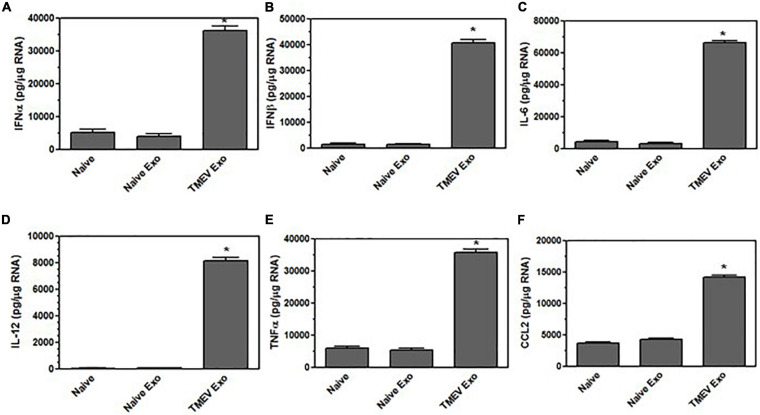
FIGURE 3.
Exosomes from TMEV-infected microglia activate bystander microglia to express type I interferons and pro-inflammatory cytokines. Exosomes were isolated from TMEV-infected microglia or uninfected (naïve) microglia (100 μg) and placed on naive microglia (1 × 10⁶) for 24 h. Microglia were lysed, RNA isolated, converted to cDNA, and analyzed by real time PCR for expression of IFNα (A), IFNβ (B), IL-6 (C), IL-12 (D), TNFα (E), and CCL2 (F). Significant difference was determined by the one way ANOVA and Bonferroni’s multiple comparison test (*p < 0.001) based on unstimulated microglia. These are representative graphs from one experiment of five independent repeated experiments.

