Fig. 2.

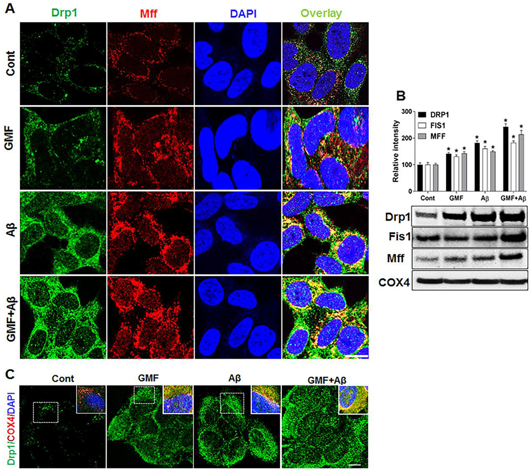
GMF increases the expression of mitochondrial fission proteins. Representative triple-labelling confocal microscopy images (a)showing staining of mitochondrial fission proteins Drp1 (green), Mff (red) and DAP1 (blue) scale bar=20 μm. Inserts are zoomed images of the boxed areas. Co-localization of fission protein Drp1 (green) along with the mitochondrial COX4 (red) staining shown in (c). Western blotting and quantitative analysis of mitochondrial fission protein, Drp1, Fis1 and Mff were performed using mitochondrial fraction from whole cell lysate. GMF treated group show higher expression of fission protein compared with control group shown in (b). Values are expressed as mean ± SE (n=3). *P<0.05 versus untreated control group.
