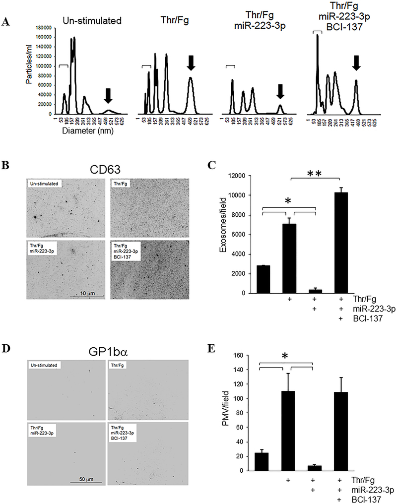
Figure 4.
Platelet transfection with miR-223–3p suppresses exosome and microvesicle release induced by thrombin/fibrinogen. Leukocyte-depleted platelets at 2 × 10⁸/ml were stimulated and transfected with 200 nM miR-223–3p mimic +/− 10 μM BCI-137 as shown for 18 hours, then platelets were collected by centrifugation and discarded. (A) Platelet releasates in the resulting supernatants were analyzed by NTA. Representative NTA tracings are shown. Brackets indicate 50–100 nm exosomes; arrows indicate ~400–550 nm diameter PMV. (B) Particles in releasates were fixed and captured on poly-L-lysine-coated coverslips, and stained with α-CD63 and α-GP1bα antibodies. Shown are the central segments of representative micrographs of exosomes imaged by epifluorescence microscopy for CD63 staining. (C) Exosomes/field were counted from full-frame micrograph images as in (B) for at least 5 fields per sample, shown as mean values after background subtraction + s.e.m. (D) Representative images of the same captured material as in (C), stained with GP1bα antibodies to label PMV. (E) PMV/field were counted from images as in (D) for at least 5 fields per sample, shown + s.e.m. *, p < 0.01; **, p < 0.02.

