FIGURE 2.
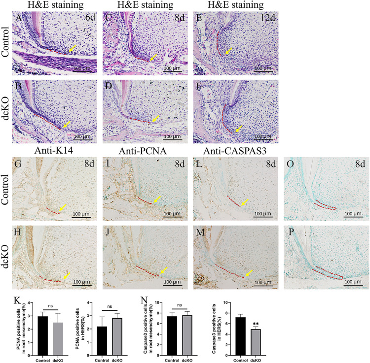
The cell proliferation and death in the persistent HERS of the dcKO molar root. (A) The H&E staining of the 1st molar root of P6 WT mouse. (B) The H&E staining of the 1st molar root of P6 dcKO mouse. (C) The H&E staining of the 1st molar root of P8 WT mouse. (D) The H&E staining of the 1st molar root of P8 dcKO mouse. (E) The H&E staining of the 1st molar root of P12 WT mouse. (F) The H&E staining of the 1st molar root of P12 dcKO mouse. (G) The K14 staining in the 1st molar root of P8 WT mouse. (H) The K14 staining in the 1st molar root of P8 dcKO mouse. (I) The PCNA staining in the 1st molar root of P8 WT mouse. (J) The PCNA staining in the 1st molar root of P8 dcKO mouse. (K) The statistical analysis of PCNA densities in the root mesenchyme and HERS. (L) The Caspase 3 staining in the 1st molar root of P8 WT mouse. (M) The Caspase 3 staining in the 1st molar root of P8 dcKO mouse. (N) The statistical analysis of Caspase 3 densities in the root mesenchyme and HERS. (O) Negative control for the anti-K14 immunostaining in the 1st molar root of P8 WT mouse. (P) Negative control for the anti-K14 immunostaining in the 1st molar root of P8 dcKO mouse. (The dashed red lines indicated the HERS; the yellow arrows delineated the end of HERS; ∗∗p < 0.01; nsp > 0.05).

