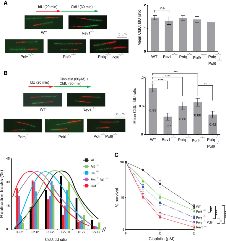
Figure 2.
Analysis of RF progression through cisplatin-induced intrastrand cross-links in wild-type (WT), Rev1−/−, Polη−/−, Polθ−/−, and Polη−/− Polθ−/− primary MEFs. (A, left) Schematic of DNA fiber assay and images of stretched DNA fibers in untreated primary WT, Rev1−/−, Polη−/−, Polθ−/−, and Polη−/− Polθ−/− MEFs. (Right) Quantification of RF progression (mean CldU:IdU ratio) in these MEFs. (B) Analyses of RF progression through cisplatin-induced intrastrand cross-links in primary WT, Rev1−/−, Polη−/−, Polθ−/−, and Polη−/− Polθ−/− MEFs. (Top left) Schematic of DNA fiber assay and representative images of stretched DNA fibers. (Top right) Quantification of RF progression through cisplatin-induced cross-links (mean CldU:IdU ratio). In A and B, quantification was done on ∼400 DNA fibers from four independent experiments. Error bars indicate the standard deviation of results of four independent experiments. Student's two-tailed t-test P-values,(**) P < 0.01, (***) P < 0.001, (****) P < 0.0001. (Bottom left) The percentage of replication tracks and the Cldu:IdU ratio measured in fibers from cisplatin-treated primary MEFs. (C) Defects in TLS through intrastrand cross-links reduce survival in cisplatin-treated primary MEFs. Error bars indicate the standard deviation from four independent experiments. Student's two-tailed t-test P-values, (*) P < 0.05, (**) P < 0.01, (***) P < 0.001, (****) P < 0.0001.

