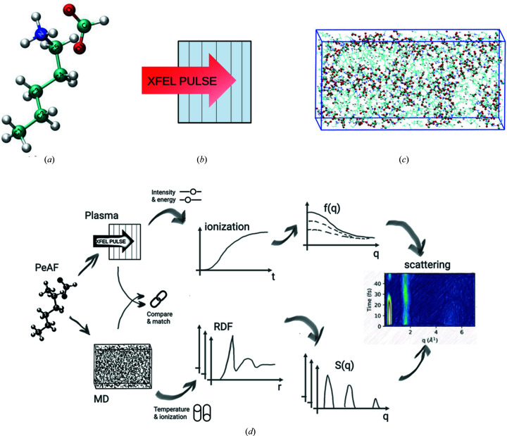
Figure 1.
(a) A snapshot of pentylammonium formate (PeAF) consisting of a pentylammonium cation and a formate anion. The molecular ions are shown in stick representation with the following colour code: cyan, blue, red and white for carbon, nitrogen, oxygen and hydrogen, respectively. (b) A schematic representation of 1D simulation geometry of plasma simulation where the homogeneous sample is divided into six distinct zones separated by black thin lines. The zones contain only information on the stoichiometry of the sample (H:15, C: 6; N:1, O:2) and no molecular structure. (c) Snapshot of a classical MD simulation box containing 300 PeAF ion pairs settled arbitrarily after initial minimization and equilibration. Colour coding as in (a). (d) Sketch of the simulation and analysis flow, combining the plasma and MD simulations, to obtain the final scattering from the liquid. X-ray beam intensity and energy are represented as ‘sliders’ and were varied to obtain the ionization dynamics and scattering form factors. Temperature and ionization in MD are represented as ‘switches’ and show different simulation scenarios for calculating the RDFs and structure factors.

