Figure 1.
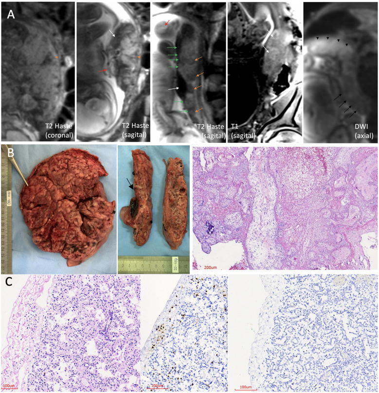
Fetal–placental magnetic resonance imaging (MRI); pathology of the placenta and lung histopathology. (A) 3.0 Tesla MRI of placenta and fetus, using two 18-channel abdominal coil, showing the umbilical cord with no evident flow, no evident signal void (red arrow), and heterogeneous placenta, with ill-defined cotyledons, with contours having fewer lobulations than usual and wedge-shape or round-shape hypointense signal areas in T2 Haste sequence (orange arrowhead and orange arrows, respectively). The placental image showed insertion in the posterior wall, with a “cake” aspect, and the uterine insertion base is smaller than usual, which gave it a “shrinkage” aspect (coronal and sagittal T2 Haste images). Altered habitual T1 and T2 placental signals with hyposignal plate in T2 along its entire length of the fetal face, appearance of a line drawn in pencil (green arrows), presence of large and dilated vessels with massive thrombosis characterized by the unusual hyperintense signal in T2, and also blood lakes between the placental tissue (areas of hypointense signal in T2 and hyperintense signal in T1, predominating in the periphery) (white arrows). Diffusion-weighted image (DWI) sequence shows higher restriction than the usual pattern, and this is more evident in the right posterolateral portion of the placenta (black arrowheads), where vascular dilation and thrombosis are more pronounced. There are also areas of necrosis characterized by the facilitation mechanism more evident in the left placental portion (black arrows). (B) Placenta pathology. Left: maternal surface is wrinkled, compact, and yellow-tan with patchy hemorrhages. Middle: cut surfaces with extensive and diffuse fibrin deposition and recent infarcts. At the level of the umbilical cord insertion, there is a large subchorionic thrombus (arrow). Right: histological section with villitis, intervillositis, and massive fibrin deposition with intervillous thrombosis. (C) Histological sections of the lung. Left: thick alveolar septa with aggregates and scattered lymphocytes in pleura (hematoxylin and eosin). Middle: immunohistochemistry showing CD3+ T lymphocytes in the pleura and alveolar septa. Right: immunohistochemistry showing no lymphocytes in a normal control lung (patient without COVID-19).

