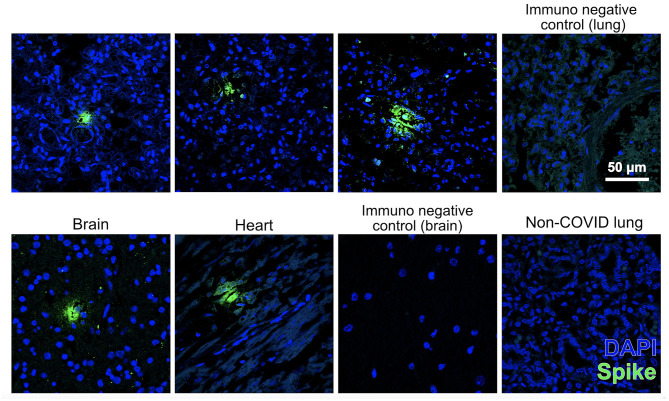
Figure 2.
Fetal tissues immunostained for SARS-CoV-2 spike protein (SP) (green) counterstained with nuclear marker DAPI (blue). Images were acquired with a confocal microscope Leica TCS SP8 using a × 63 objective lens. Scale bar for all pictures is 50 μm. Top pictures: pulmonary immunodetection of SP (the three on the left) and negative control of immunofluorescence without primary antibody (right) in the present case. Lower images: immunodetection of SP in the brain and heart of this case (the two on the left), negative control immunofluorescence without primary antibody in a section of the brain, and lack of immunodetection of SP in a non-COVID fetal lung (right).

