
Figure 1.
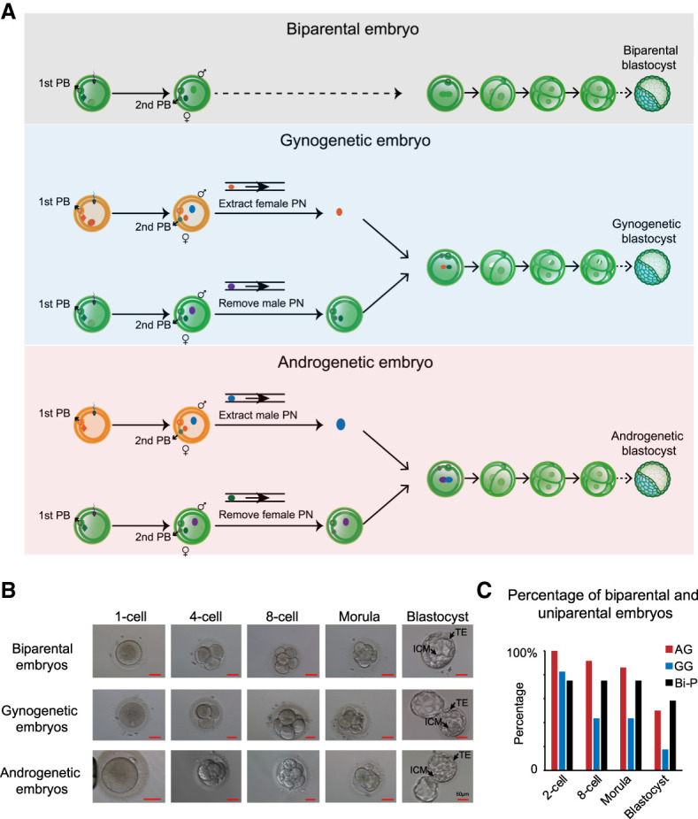
Generation of human diploid AG and GG embryos. (A) Schematic illustration of human diploid Bi-P, GG, and AG embryo generation. Briefly, following ICSI, two oocytes were fertilized. Four to six hours later, female pronuclei were identified based on their proximity to the second (2nd) polar bodies and smaller size compared with male pronuclei. The female pronucleus from individual zygotes was removed to generate haploid androgenotes. The male pronucleus from the donor androgenote was then extracted and mixed with Sendai virus and transferred into the recipient androgenote, leading to the formation of a diploid androgenote for subsequent development into a blastocyst. GG embryos were produced with the same method. (B) The development of Bi-P and uniparental embryos to blastocysts after pronuclei transfer. The uniparental embryos developed blastocysts with identifiable TE and ICM, the same as the Bi-P embryos in vitro. (C) The development ratios of Bi-P, GG, and AG embryos.