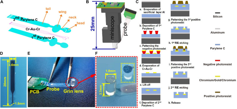
FIGURE 1.
(A) The design of the probe; (B) the concept diagram of the modified system for simultaneously observing Ca2+ and cortical electrophysiology; (C) the microfabrication process of the electrode probe from the cross-sectional view; (D) the fabricated probe; (E) the assembled probe and GRIN lens; and (F) the zoom-in view of the imaging and recording regions of the electrode probe as red dotted circles in panel (E). The inset circled by a dotted box is another type of probe designed in the experiment for imaging comparison.

