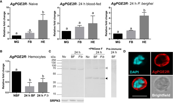
Figure 1.
Characterization of a prostaglandin E2 receptor (PGE2R) in Anopheles gambiae. (A) AgPGE2R gene expression was examined by qRT-PCR in midgut (MG), fat body (FB), and perfused hemocytes (HE) under naïve (NV), blood-fed (24 h BF), or P. berghei-infected (24 h P.b) conditions. (B) AgPGE2R expression was more closely examined in hemocytes and compared across physiological conditions. For both (A, B), letters indicate statistically significant differences (P < 0.05) when analyzed by a one-way ANOVA followed by a Tukey’s multiple comparison test using GraphPad Prism 7.0. Bars represent the mean ± SE of either three or four independent biological replicates. (C) Perfused hemolymph from naïve (NV), blood-fed (BF), or P. berghei infected (P.b) mosquitoes were examined by Western blot analysis. Specific bands were detected corresponding to a glycosylated AgPGE2R product (bands at ~70 kDa, open arrowhead), or in which the receptor underwent deglycosylation with PNGase F treatment (~46 kDa, black arrowhead). The AgPGE2R was detected using a rabbit antibody (1:1000) directed against intracellular loop 3 (ICL3). No bands were detected in a 24 h BF hemolymph sample incubated with pre-immune serum. Serpin 3 (SRPN3) was used as a protein loading control. (D) Immunofluorescence assays were performed on perfused hemocytes using a rabbit polyclonal antibody (1:500) against extracellular loop 2 (ECL2), revealing that PGE2R is localized to oenocytoid immune cell populations (scale bar, 5 µm).

