FIGURE 3.

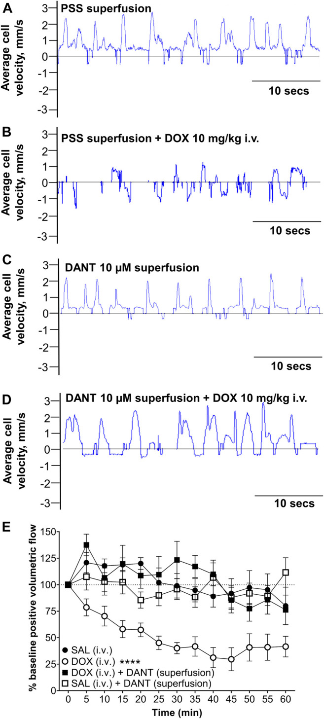
Superfusion of DANT preserves in vivo lymph flow in mesenteric LVs of anesthetized rats infused with DOX. Frame-by-frame cell tracking (300 fps video capture) measures the distance travelled by a cell in lymph fluid as a function of time to enable estimation of cell velocity. (A) Upward deflections indicate positive (distal-to-proximal) cell velocity in control LVs superfused with PSS. (B) Infusion of DOX (10 mg/kg i.v.) reduced average lymph cell velocity to cause “lymphostasis,” which is evident by lower amplitude upward deflections interspersed with downward deflections indicating back flow. (C) Superfusion of the mesenteric LVs with DANT (10 μmol/L) had no effect on average lymph cell velocity. (D) Additionally, lymph cell velocity was preserved in mesenteric LVs superfused with DANT prior to infusion of DOX (10 mg/kg i.v.). (E): Compared with an equi-volume infusion of saline (SAL–filled circles; n = 6), systemic administration of DOX (DOX; open circles; n = 7) markedly reduced positive volumetric flow in rat mesenteric LVs. Addition of DANT to the superfusate 20 min prior to and continuously after- systemic administration of DOX preserved lymph flow (DOX + DANT; filled squares; n = 7). Positive volumetric lymph flow in mesenteric LVs superfused with DANT was stable in animals infused with SAL (SAL + DANT; open squares; n = 7). Data reported as mean ± S.E.M. and analyzed using two-way ANOVA (F (3.204, 15.13) = 2.435). **** significant from SAL, DOX + DANT, and SAL + DANT; p < 0.0001.
