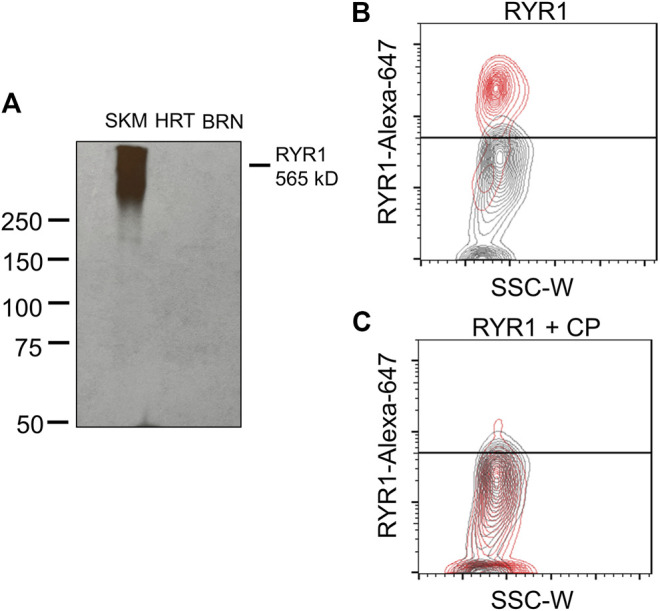
FIGURE 5.

RYR1 antibody selectivity. (A) Western blot shows positive detection of RYR1 (∼565 kD) in rat skeletal muscle lysate and no detection in rat heart or brain lysate. (B,C) Contour plots of RYR1 antibody testing. (B) Red population represents positive detection of RYR1 subtype in male LMCs. Grey population represents the LMC suspension containing no primary antibody. (C) No detection in male LMCs after co-incubation with competing peptide (CP; 1:50 dilution) for the RYR1 antibody. Data representative of three isolations for each sex (n = 6).
