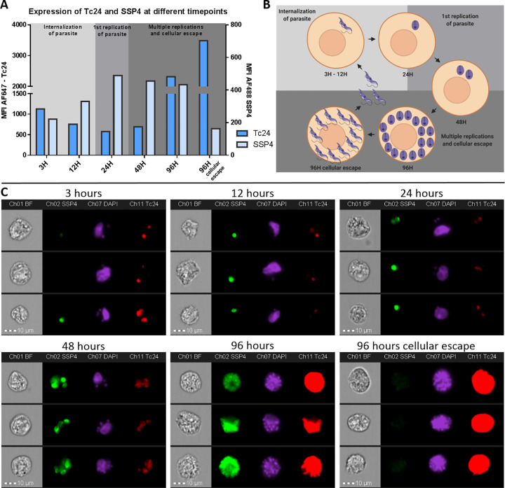
Fig 7. Imaging Flow Cytometry reveals a change in expression of Tc24 and SSP4 during different infection timepoints of T. cruzi.
VERO cells were infected with T. cruzi and 3 hrs, 6 hrs, 12 hrs, 24 hrs, 48 hrs and 96 hrs after start infection fixed and permeabilized. VERO cells were then stained for Tc24 and SSP4. Nuclei were stained using DAPI. A) The MFI of Tc24 and SSP4 from the total population of T. cruzi—infected VERO cells was plotted in a histogram. The left y-axis depicts the MFI for Tc24 and the right y-axis depicts the MFI for SSP4. B) A schematic representation of the transformation of T. cruzi trypomastigotes to amastigotes and back to trypomastigotes in the VERO host cell is shown. Prepared using Biorender.com C) Image gallery of three events of each infection time point representing the complete population. Presented are brightfield, Tc24 (red), SSP4 (green) and DAPI (magenta).

