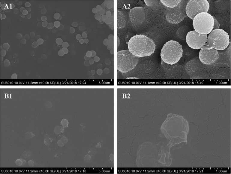
FIGURE 4.
Scanning electron microscopy images of biofilms formed by Staphylococcus epidermidis following 12 h of treatment with 16 μg/mL aloe-emodin. Treatment without (A) and with aloe-emodin (B). A2 and B2 show higher magnifications than A1 and B1, respectively. Scale bars of 5 μm and 1 μm are shown at the bottom right of the panels marked (A) and (B), respectively, with each scale bar equally divided into 10 units.

