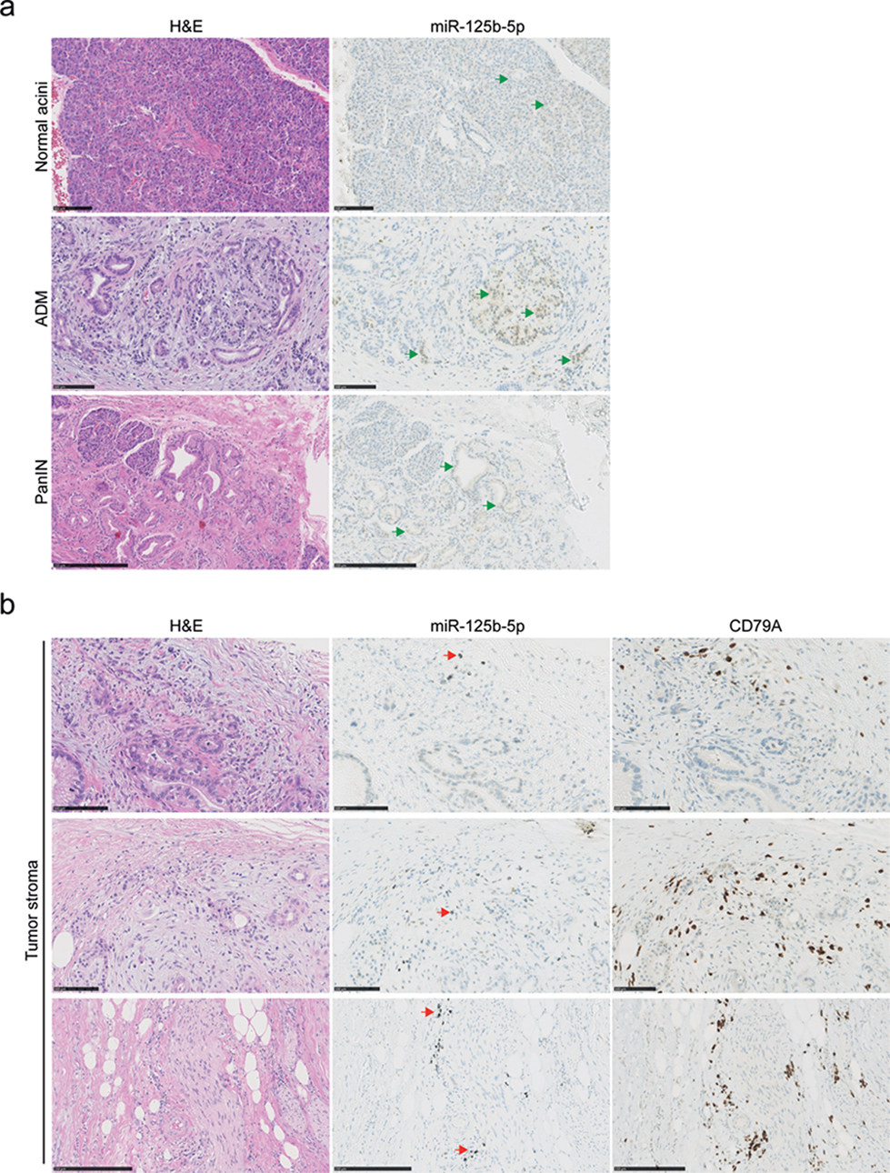
Figure 3.

MiR-125b-5p detection by in situ hybridization in pancreatic cancer tissues. Serial FFPE tissue sections were stained with H&E or a DIG labeled miR-125b-5p probe. MiR-125b-5p expression is detected by silver staining resulting in brown/black coloring, whereas nuclei are stained with hematoxylin in blue. (a) Representative images of low expression of miR-125b-5p, (green arrows), in untransformed pancreatic acinar cells and cells that underwent acinar to ductal metaplasia (ADM) or pancreatic intraepithelial neoplasia (PanIN). Scale bars are 100 μm in the normal acini and ADM; scale bar is 250 μm in the PanIN. (b) Representative images of cells in the tumor stroma with high expression of miR-125b-5p (red arrows). Corresponding tissue sections are stained with H&E or the B-lymphocyte lineage marker CD79A antibody by immunohistochemistry (brown). Scale bars are 100 μm in the top two rows; scale bars are 250 μm in the bottom row.
