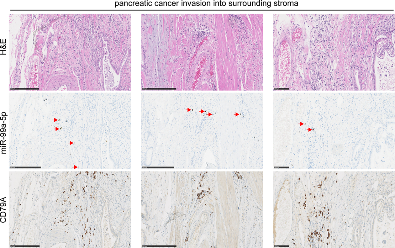
Figure 4.

MiR-99a-5p detection by in situ hybridization in pancreatic cancer tissues. Serial FFPE tissue sections were stained with H&E a DIG labeled miR-99a-5p probe, or the B-lymphocyte lineage marker CD79A antibody by immunohistochemistry (brown). Red arrows indicate miR-99a-5p expression, detected with silver staining resulting in brown/black coloring, whereas cell nuclei are stained with hematoxylin in blue. Scale bars are 250 μm in the first two columns; scale bar is 100 μm in the right-most column.
