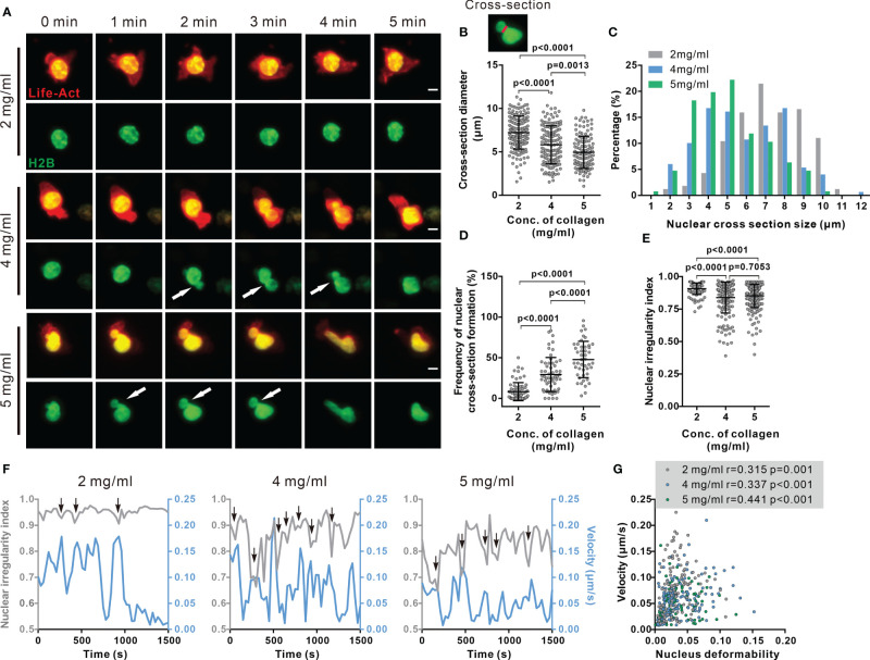
Figure 3.
Nuclear deformation is correlated with migration velocity in dense collagen matrices. (A–E) Nuclear deformation in CTLs is enhanced upon the increase in matrix density. CTLs transfected with Histone 2B-GFP (green) and LifeAct-mRuby (red) were embedded in collagen. Migration was visualized using light-sheet microscopy (20× objective) at 37°C every 30 sec for 30 min. Exemplary cells are shown in (A). Cross-sections are pointed by the arrowheads. Scale bars are 5 µm. Quantification of cross-section diameter, their distribution and duration is shown in (B) (indicated as red-line in the inset), (C, D). The nuclear irregularity index of the cells in (A–D) is shown in (E). Results are presented as Mean ± SD for (B, D, E). All results are from 3 donors. The Mann-Whitney test was used for statistical significance. (F, G) Migration velocity of CTLs is positively correlated with nuclear deformation. NII (sphericity) and velocity as function of time in exemplary cells are shown in (F). Correlation between velocity and nuclear deformability is shown in (G). One dot represents one cell. Correlation coefficient r is analyzed with Spearman’s correlation. All results are from 3 donors.

