Figure 1.
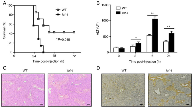
N-3 PUFAs exacerbate APAP-induced liver damage. (A) APAP (600 mg/kg) was injected into WT and fat-1 transgenic mice (n=7), and the survival rate of mice was observed (P=0.015). (B-D) WT and fat-1 transgenic mice (n=5) were intraperitoneally injected with APAP (400 mg/kg). (B) Serum ALT activities at different time points were measured. (C) Histological analysis of mouse livers was performed by hematoxylin and eosin staining 24 h post APAP injection. Scale bar, 100 µm. (D) TUNEL staining was performed on paraffin-embedded liver sections 24 h after APAP injection to mark apoptotic cells. Scale bar, 100 µm. *P<0.05, **P<0.01. N-3 PUFAs, omega-3 polyunsaturated fatty acids; APAP, acetaminophen; WT, wild-type; ALP, alanine aminotransferase.

