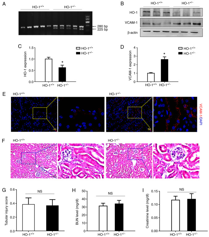
Figure 1.
HO-1+/− knockdown mice exhibit elevated expression levels of VCAM-1 without changes in renal structure and function. (A) Genotype identification of HO+/+ and HO+/− mice by agarose gel electrophoresis. (B) Western blot analysis of the expression levels of HO-1, VCAM-1 and β-actin proteins in the HO-1+/− vs. those in the wild-type mice. (C and D) Densitometric-based quantification of the western blot analysis results shown in panel B for (C) HO-1 and (D) VCAM-1 proteins using ImageJ software. Densitometry values are expressed as the mean ± SD (n=3). *P<0.05 vs. HO-1+/+. (E) Immunohistochemical staining of VCAM-1-expressing cells in the kidneys of the HO-1+/+ and HO-1+/− mice. (F) Representative images of H&E-stained sections of renal tissue in the HO-1+/+ and HO-1+/− mice. (G) Tissue injury was assessed by using the scoring scale from 0 to 5 points (n=3). (H) Serum creatinine concentration in the HO-1+/+ and HO-1+/− mice (n=3). (I) Serum BUN concentration in the HO-1+/+ and HO-1+/− mice (n=3). HO-1, heme oxygenase-1; VCAM-1, vascular cell adhesion molecule-1; BUN, blood urea nitrogen.

