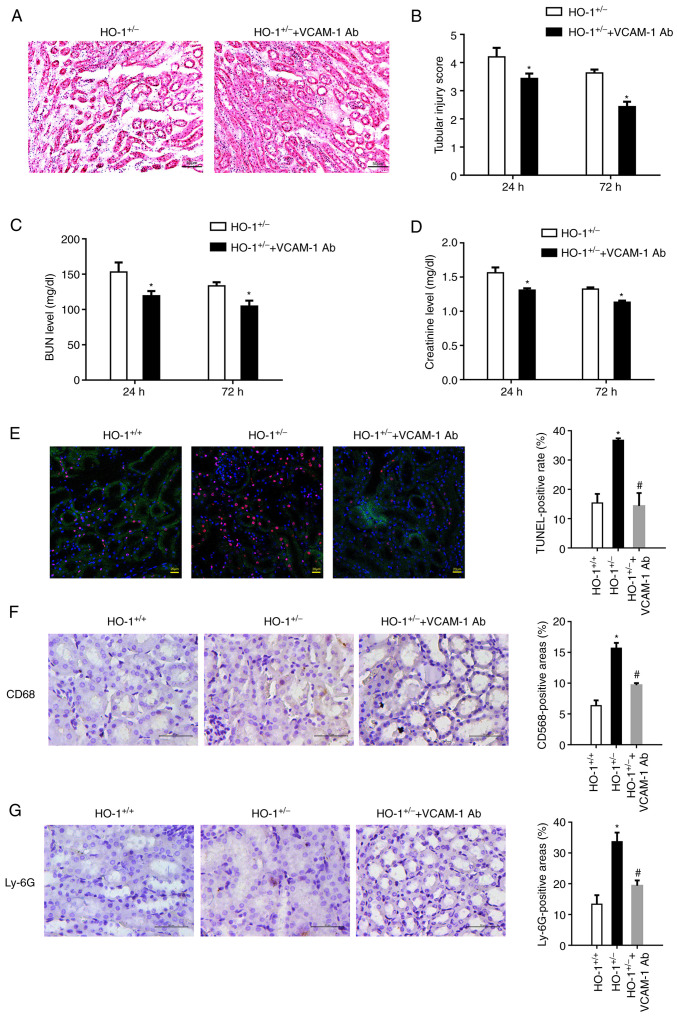
Figure 3.
VCAM-1 blocking alleviates renal IRI. VCAM-1 antibody was infused into the HO-1+/− knockdown mice through the tail vein to block VCAM-1 expression on the vascular endothelium. (A) Representative images of H&E-stained sections of renal tissue showing its morphology. (B) Extent of the kidney tissue injury was assessed using the 0 to 5-point scoring system (n=3, *P<0.05 vs. HO-1+/−). (C) Serum BUN concentration at 24 and 72 h post-IRI in the HO-1+/+ and HO-1+/− mice (n=3, *P<0.05 vs. HO-1+/−). (D) Serum creatinine concentration at 24 and 72 h post-IRI in the HO-1+/+ and HO-1+/− mice (n=3, *P<0.05 vs. HO-1+/−). (E) Cell death upon IRI was measured using TUNEL assay. The TUNEL-positive rate was analyzed using ImageJ software in the HO-1+/+, HO-1+/− and HO-1+/− + VCAM-1 Ab groups (*P<0.05 vs. HO-1+/+; #P<0.05 vs. HO-1+/−). (F) Immunohistochemical staining and quantification analysis of CD68-expressing cells in mouse kidneys in the HO-1+/+, HO-1+/− and HO-1+/− + VCAM-1 Ab groups (*P<0.05 vs. HO-1+/+; #P<0.05 vs. HO-1+/−). (G) Immunohistochemical staining and quantification of Ly-6G-expressing cells in mouse kidneys of the HO-1+/+, HO-1+/− and HO-1+/− + VCAM-1 Ab groups (*P<0.05 vs. HO-1+/+; #P<0.05 vs. HO-1+/−). IRI, ischemia-reperfusion injury; HO-1, heme oxygenase-1; VCAM-1, vascular cell adhesion molecule-1; BUN, blood urea nitrogen; Ab, antibody.

