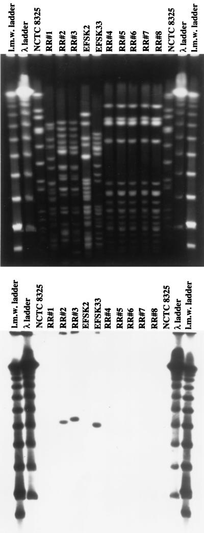
FIG. 1.
PFGE patterns of isolates recovered from the PD patient. Chromosomal DNA was prepared, SmaI restricted, and separated by PFGE (see Materials and Methods). The bottom panel shows results of hybridization with a probe for vanA. E. faecium strains EFSK2 (vanB) and EFSK33 (vanA) were used as controls. l.m.w., low molecular weight.

