Fig. 2.

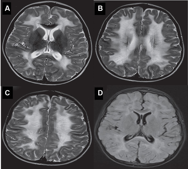
( A–C ) Axial T2WI showing diffuse periventricular and lobar white matter hyperintensity with a somewhat striated appearance and Axial FLAIR image showing partial inversion of the frontal and parietal white matter lesions noted in ( D ).

( A–C ) Axial T2WI showing diffuse periventricular and lobar white matter hyperintensity with a somewhat striated appearance and Axial FLAIR image showing partial inversion of the frontal and parietal white matter lesions noted in ( D ).