FIG. 4.
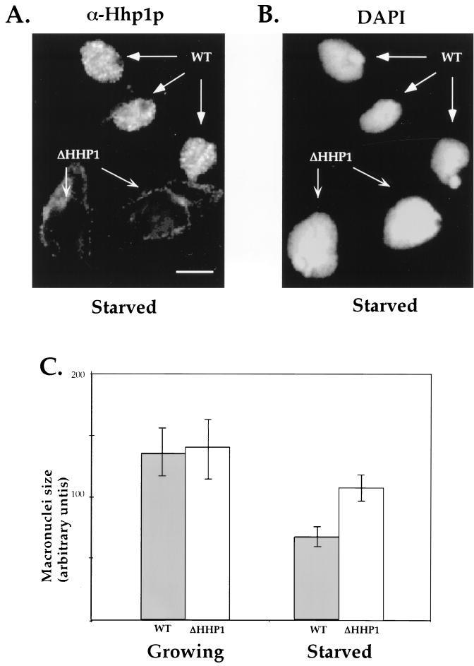
ΔHHP1 cells display abnormally large macronuclei upon starvation. Wild-type (WT) and HHP1 knockout (ΔHHP1) cells were starved in 10 mM Tris for 24 h, mixed at a 1:1 ratio, and stained in situ with α-Hhp1p antibodies (A) and DAPI (B). As expected, a punctate Hhp1p staining pattern was observed in the macronuclei of wild-type cells, while no staining was detected in ΔHHP1 cells (see arrows in panel A). Note the relatively larger size of macronuclei in the ΔHHP1 cells. Bar, 10 μm. (C) The DAPI-stained cross-sectional areas of 100 to 200 macronuclei from growing or starved wild-type or ΔHHP1 cells were measured; the nuclear size is presented in arbitrary units. The mean and standard error values for three independent experiments are shown. Note that no significant difference between the macronuclei of wild-type and ΔHHP1 cells is detected under growing conditions, while an approximately 1.5-fold difference is detected between cell strains after starvation.

