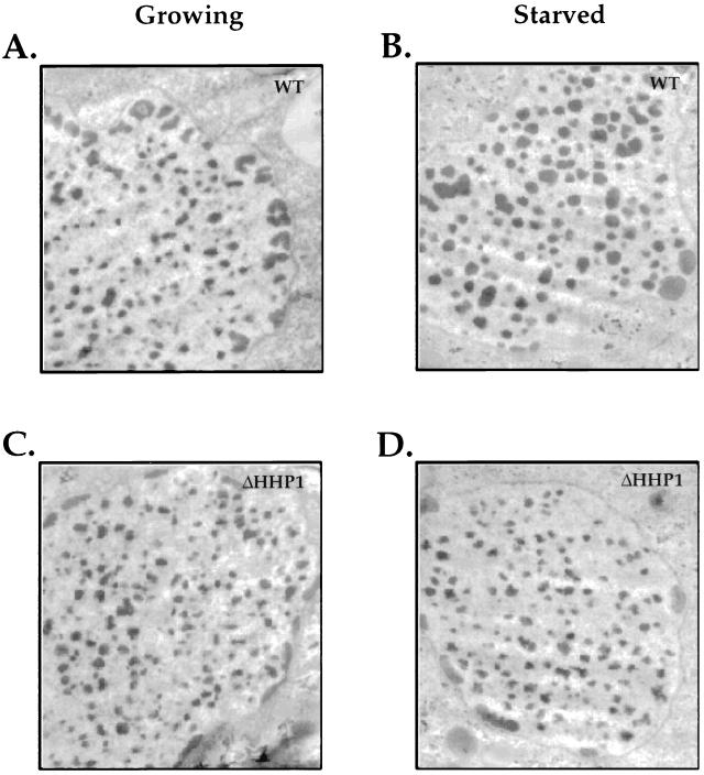
FIG. 5.
Ultrastructural analyses of chromatin bodies in macronuclei of wild-type and ΔHHP1 cells. Growing (A and C) or starved (B and D) wild-type (A and B) or ΔHHP1 (C and D) cells were fixed and processed for ultrastructural analysis by transmission electron microscopy. Note the difference in size between the chromatin bodies of the wild-type and ΔHHP1 cells, particularly in the starved cells. No consistent differences in the size or morphology of the multiple, peripherally located nucleoli were observed between the strains.

