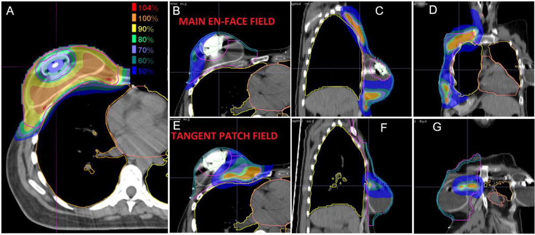
Figure 4:
Axial (A, B, E), sagittal (C, F), and coronal (D, G) 50% color wash images demonstrating a two proton field, multi field optimization (MFO) plan avoiding delivery through the magnetic expander port of the reconstructed breast while achieving comprehensive target coverage. Individual dose deposition profiles from the en face field (B-D) and the more tangential beam angle (D-F) are displayed.

