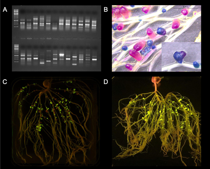
FIGURE 2.
Visualization of some methods for assessing rhizobial competitiveness for nodulation. (A) Strain-specific genomic fingerprints: ERIC-PCR from same plant nodule isolates from a trapping assay using faba bean as a host; (B) Sequential double staining to detect gusA and celB: Pea roots were sequentially double-stained with Magenta-GlcA and X-Gal after thermal treatment, resulting in pink nodules formed by UPM791gusA (gusA constitutively expressed) and blue nodules formed by Rlv3841celB (celB constitutively expressed); (C) Fluorescent proteins: Rlv3841 labeled with mini-Tn7 J23104 GFP or mCherry, respectively; and (D) NGS of synthetic DNA fragments: Example of pea roots grown in non-sterile soil and exposed to a blue-light transilluminator. Tagged rhizobia, expressing GFP under PsnifH control, lead to fluorescent nodules, while indigenous rhizobia do not. Photo credit: (A,B,D) Marcela Mendoza-Suárez; (C) Laura Clark.

