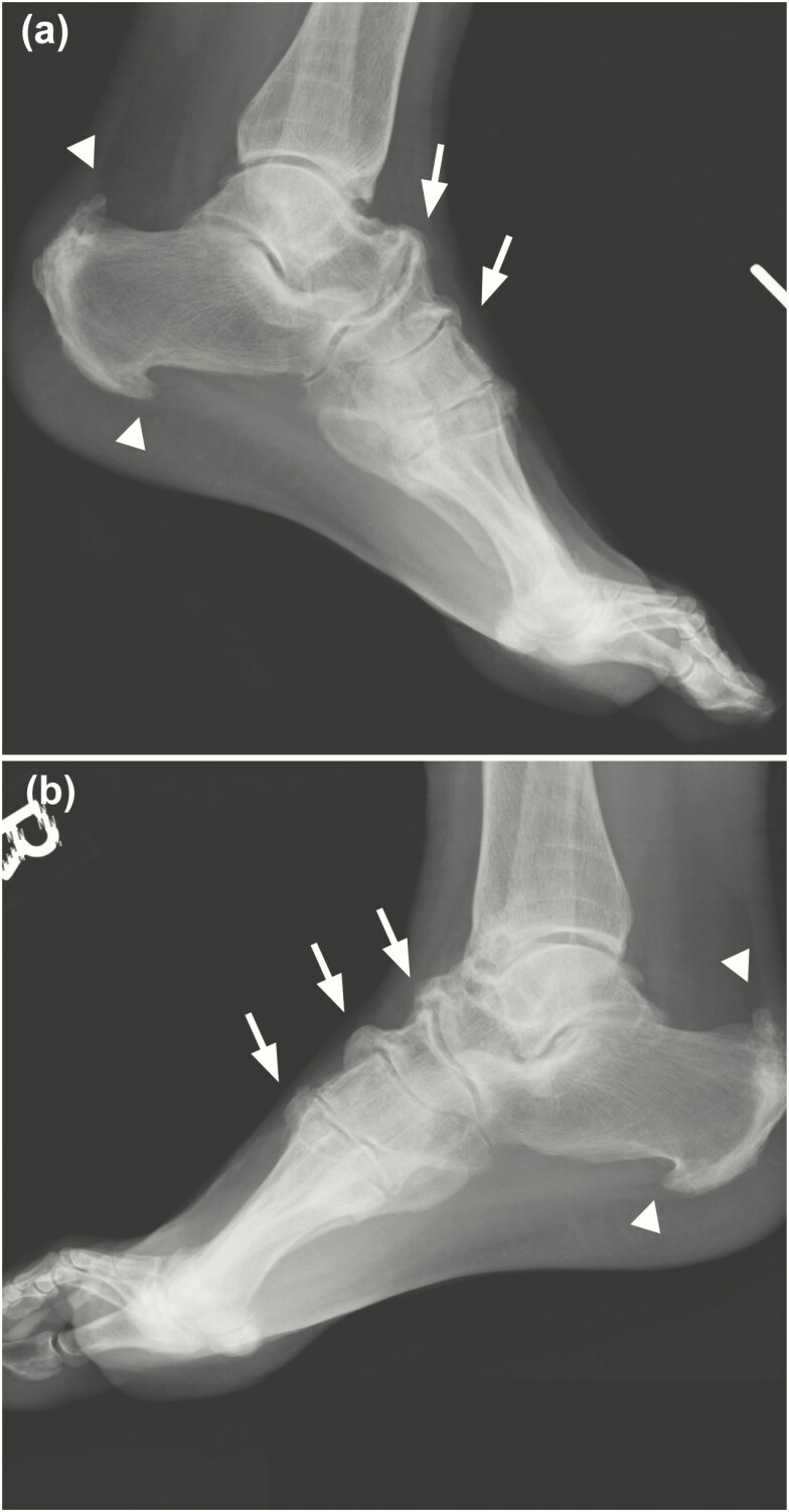
Figure 5.
Radiographic imaging of the feet of a 59-year-old man with X-linked hypophosphatemia, representative of study participants. Growth arrest lines visible on the distal portions of the tibia and lateral A, left and B, right x-rays show prominent symmetrical enthesophyte formation involving the plantar and posterior aspects of calcanei, bilaterally (arrowheads). Enthesophytes present at the talonavicular, cuneonavicular, and metatarsal cuneiform ligaments attachment to the dorsal foot (arrows).

