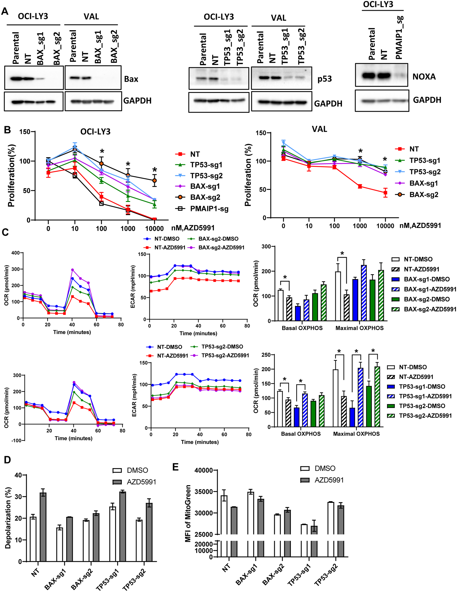
Figure 4. Loss of TP53 and BAX mediates resistance to Mcl-1 inhibition.

(A) OCI-LY3 and VAL cells were electroporated with RNP single sgRNA/Cas9 constructs targeting TP53, BAX, PMAIP1 or nontargeting sgRNA (NT). Whole protein lysates were subjected to immunoblotting. (B) Genetically manipulated cells were treated with the indicated concentrations of AZD5991 and proliferation was measured in the MTS assay. % viable cells, after normalization to untreated control, were fit using nonlinear regression analyses. Data are Mean±SE. * - p<0.05 vs. sgTP53/BAX vs. NT control (C-E) Genetically manipulated VAL cells were treated with 1 μM AZD5991 or vehicle control for 24 h and subjected to Seahorse analysis (C), analysis of mitochondrial depolarization (JC-1; D) and mitochondrial mass (Mitobright Green; E).
