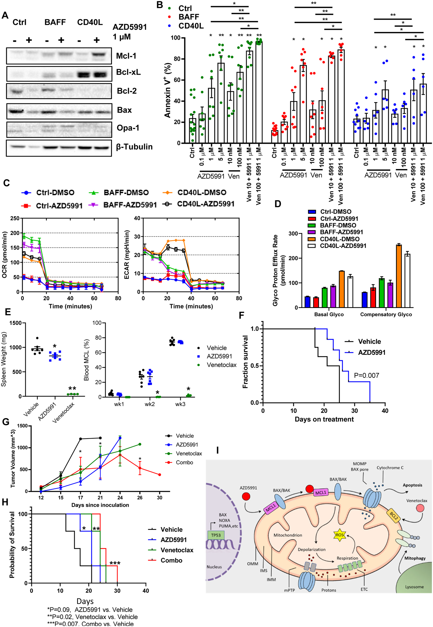
Figure 6. Mcl-1 inhibition overcomes stromal rescue of the primary MCL cells and demonstrates efficacy in vivo.

(A-B) Primary MCL cells (4_individual patient samples, in duplicates) were co-cultured with BAFF-, CD40L-expressing or control stroma for 24 h and then treated with the indicated drugs for additional 24 h. Cells were assayed for apoptosis and protein lysates were analyzed by immunoblotting. (C, D) Primary MCL cells were cultured on stroma as indicated, treated with 1 μM AZD5991 for 24 hours and subjected to Seahorse respiration assay. (E-F) MCL PDX mice received treatment with AZD5991 (N=8), venetoclax (N=4) or vehicle control (N=8) as described in the methods. Treatment was begun upon detection of the circulating MCL cells in the peripheral blood. Spleen weight was measured at the time of sacrifice. % MCL involvement and survival are shown since start of treatment. (G-H) Mice xenografted with OCI-LY3 cells were treated with AZD5991, venetoclax, the combination or vehicle control as discussed in the methods (N=4 each). Tumor volume and survival are shown. X axis represents days since tumor inoculation. * - p<0.05 each treated versus untreated tumor. (I) Pharmacologic targeting Mcl-1 induces mitochondrial dysfunction and apoptosis in B-cell lymphoma cells in TP53- and BAX-dependent manner.
