. 2021 Jun 29;27(17):4859–4869. doi: 10.1158/1078-0432.CCR-21-0851

Figure 3.

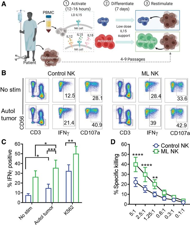
Figure 3. Memory-like NK cells from patients with advanced melanoma exhibit enhanced ability to control autologous tumor. A, PBMC containing patient NK cells were preactivated as indicated in Fig. 2A. Melanoma cell lines generated from metastatic tissue were used as target cells for the functional assays. B, Representative flow cytometry plots of control and ML NK cells from a patient with advanced melanoma stimulated with autologous tumor. Numbers indicate percentage of positive cells. ML NK cells (green) from patients with melanoma patients display superior ability to produce IFNγ upon stimulation with autologous tumor (5:1 E:T ratio) compared with control NK cells (blue; C). D, ML NK cells from patients with advanced melanoma exhibit superior ability to kill autologous melanoma targets compared with control NK cells at different E:T ratios. Killing was evaluated in a standard 4-hours 51Cr release assay. Bars represent mean ± SEM from all patients. Two-way ANOVA with Tukey post hoc analysis. *, P < 0.05; **, P < 0.01; ***, P < 0.001; ****, P < 0.0001. n = 7.

Memory-like NK cells from patients with advanced melanoma exhibit enhanced ability to control autologous tumor. A, PBMC containing patient NK cells were preactivated as indicated in Fig. 2A. Melanoma cell lines generated from metastatic tissue were used as target cells for the functional assays. B, Representative flow cytometry plots of control and ML NK cells from a patient with advanced melanoma stimulated with autologous tumor. Numbers indicate percentage of positive cells. ML NK cells (green) from patients with melanoma patients display superior ability to produce IFNγ upon stimulation with autologous tumor (5:1 E:T ratio) compared with control NK cells (blue; C). D, ML NK cells from patients with advanced melanoma exhibit superior ability to kill autologous melanoma targets compared with control NK cells at different E:T ratios. Killing was evaluated in a standard 4-hours 51Cr release assay. Bars represent mean ± SEM from all patients. Two-way ANOVA with Tukey post hoc analysis. *, P < 0.05; **, P < 0.01; ***, P < 0.001; ****, P < 0.0001. n = 7.