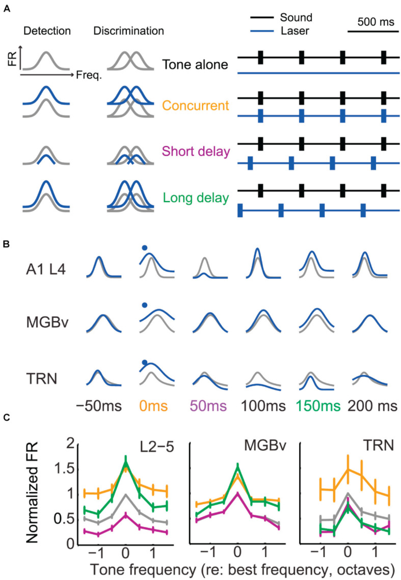
FIGURE 4.
Corticothalamic gain control on frequency tuning curves of A1, MGBv, and TRN neurons. (A) Schematic of corticothalamic modulation on the turning curves of A1 cortical neurons and optogenetic laser stimulation paradigm. Lemniscal layer VI corticothalamic neurons were activated by expressing ChR2 in A1 bilaterally in Ntsr1-Cre mice and using pulses of blue laser light. Depending on the duration of the sound stimulation delay following laser activation of corticothalamic neurons in layer VI, the tuning curves were predicted to be modulated distinctively and to define tone detection and discrimination behaviors. The animals were trained to detect or discriminate sounds using an avoidance task. (B) Representative modulation effect on the tuning curves of A1, MGBv, and TRN neurons (gray, tone-alone; blue, tone-and-laser). Sound-evoked responses enhanced in A1 and MGBv neurons for the concurrent stimulation of tones and laser (orange). For the tone presentation with a short or long delay following the corticothalamic stimulation (purple or green), corticothalamic modulation effects differed in A1, MGBv, and TRN neurons, showing enhancement for one station while showing suppression for the others. (C) Tone-evoked firing rates (mean ± SEM) were normalized and compared to the values evoked for the BF of the tone-alone condition. When tone presentation and corticothalamic stimulation were concurrent, all A1, MGBv, and TRN neurons increased firing rates (paired t test, p < 0.05). When tones were presented with a short delay following laser stimulation, the firing rates decreased in A1 and TRN neurons (p < 0.05) but no change was found in MGBv neurons (p > 0.05). For the long delayed condition, A1 and MGBv neurons increased firing rates (p < 0.05), whereas TRN neurons reduced it (p < 0.05) (Adapted and modified with permission from Guo et al., 2017; Figures 4B, 5D,E).

