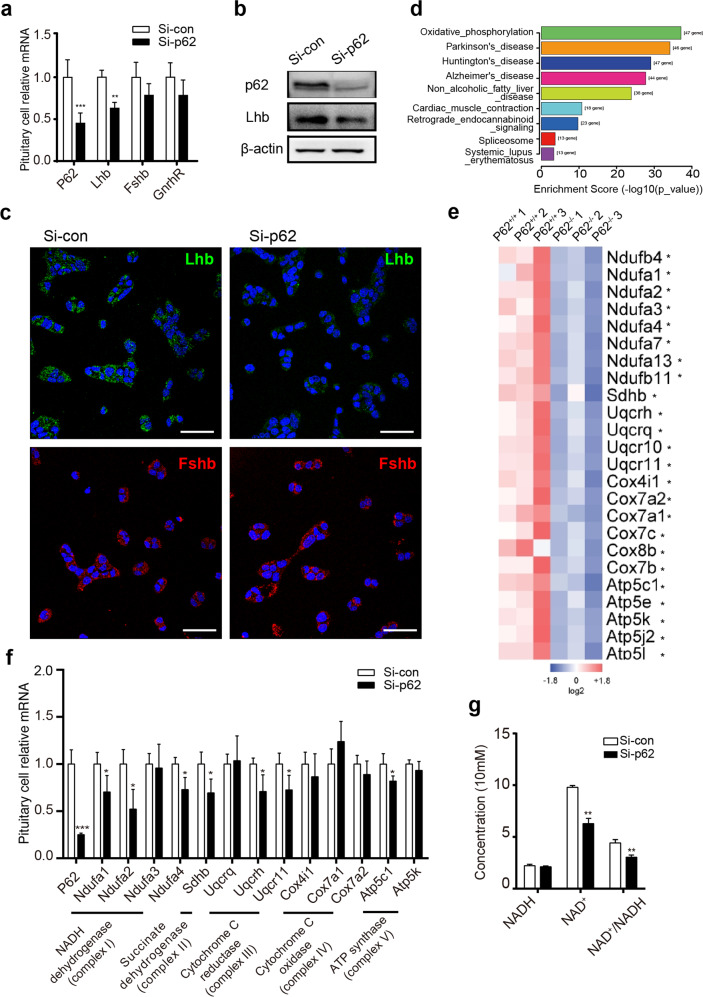
Fig. 4. P62 deficiency leads to downregulated LH and oxidative phosphorylation (OXPHOS) in pituitary gonadotropin cells.
a–c In vitro experiments in which the mouse pituitary gonadotroph LβT2 cell line was transfected with control/p62 siRNA for 48 h. a RT-PCR detection of p62, Lhb, Fshb, and GnRHR mRNA, n = 4–6. b WB detection of silenced p62 expression and LH protein levels. c Immunofluorescence detection of LH (green) and FSH (red) protein expression in LβT2 cells. DAPI in blue, scale bar: 20 μm. d The significant changed pathways in p62−/− mouse pituitaries enriched by KEGG analysis. e Differentially expressed pituitary OXPHOS markers between p62−/− and p62+/+ mice shown as a heatmap from RNA-Seq of young female mice, n = 3. f Relative mRNA expression levels of OXPHOS markers (mitochondrial complex I: Ndufa1–4; complex II: Sdhb; complex III: Uqcrq, Uqcrh, Uqcr11; complex IV: cox4i1, cox7a1, cox7a2; complex V: atp5c1, atp5k) with p62 siRNA treatment, n = 4. g NADH, NAD+ concentration and NAD+ /NADH ratio measurement, n = 4. All data are presented as the mean ± SD. Student’s t-test, *P < 0.05; **P < 0.01; ***P < 0.001.

