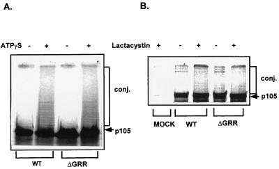
FIG. 3.
Conjugation of WT p105 and p105-ΔGRR in vitro (A) and in vivo (B). (A) In vitro-translated WT and ΔGRR 35S-labeled p105s were incubated in the presence of HeLa cytosolic extract, ATPγS (as indicated), ubiquitin, and UbAl, as described in Materials and Methods. Reaction mixtures were resolved via SDS-PAGE, and conjugates were visualized by exposure to a phosphorimager screen. (B) COS-7 cells were transiently transfected with the pCI-neo vector (lane MOCK) or with the same vector containing the cDNAs for WT or ΔGRR p105. Transfected cells were incubated in the presence of [35S]methionine and in the presence or absence of clasto-lactacystin β-lactone as described in Materials and Methods. Following labeling, proteins were immunoprecipitated with anti-p50 antibody, resolved via SDS-PAGE, and visualized by phosphorimaging as described in Materials and Methods. Conj., ubiquitin conjugates.

