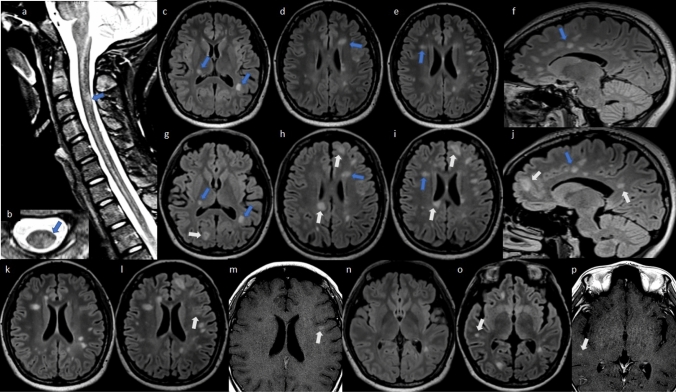
Fig. 3.
Serial 3 T MRI scans in case 3. Baseline scan: sagittal STIR (a) and axial T2 (b) of the cervical spinal cord and axial (c–e, k, n) and sagittal (f) T2 FLAIR MRI of the brain obtained 70 days prior to onset of new neurologic symptoms, showing multiple T2 hyperintense demyelinating lesions in the brain and one lesion in the spinal cord (blue arrows). Post-vaccination scan: axial (g–i, l, o) and sagittal (j) T2 FLAIR of the brain 6 days after new neurologic symptom onset and 7 days after COVID-19 vaccination, showing multiple new T2 hyperintense lesions (white arrows: new lesions, blue arrows: old lesions). T1 post-gadolinium axial images (m, p) show enhancement (white arrows) of two of the new FLAIR lesions. Spinal cord imaging was not obtained post vaccination due to the isolated visual symptoms

