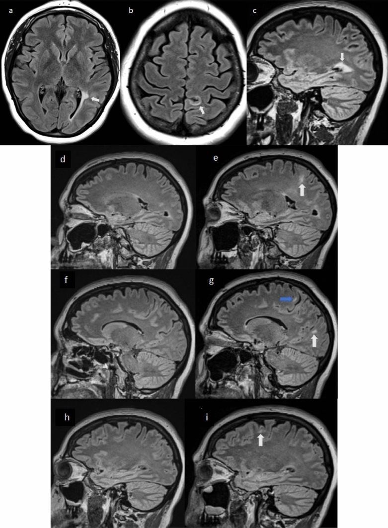
Fig. 7.
Serial 3 T MRI brain scans in case 7. Baseline (2 years prior to COVID-19 vaccination): T2 FLAIR of the brain (a–c) show three typical MS lesions (white arrows). Sagittal T2 FLAIR brain scans (d, f, h), also obtained at baseline, without any lesions. Post-vaccination: sagittal T2 FLAIR (e, g, i) obtained at the time of new neurologic symptoms following COVID-19 vaccination. Note three new lesions with typical multiple sclerosis morphology (white arrows). Also note that one of the lesions seen on image g (blue arrow), is an extension of the same lesion seen on image e

