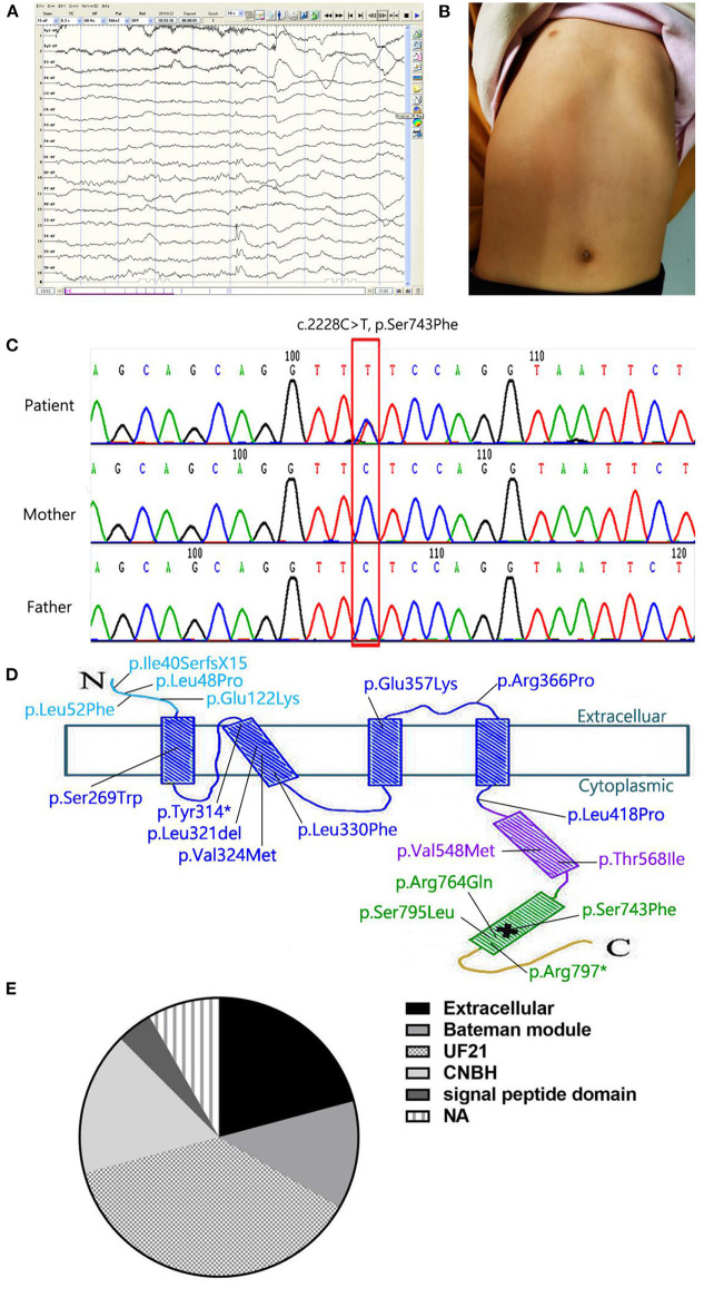
Figure 1.
(A) The EEG monitoring showed the release of sharp-slow and spinous-slow waves in the left posterior temporal region and the right middle posterior temporal regions. (B) Patient's picture showing a funnel-shaped chest. (C) Partial CNNM2 electropherograms of the patient and her parents. In the electropherograms, the variant is indicated by a red box and the changes in nucleotide and resulting effects on the protein are shown. (D) Localization of the variant in the secondary structure of CNNM2. The N-terminal extracellular domain and the transmembrane domain are in light blue and dark blue respectively. The CBS domain is in purple, the CNBH domain is in green, and the unstructured C-terminus is yellow. *means stop codon. The location of pathological variant is indicated by a cross. (E) variant domains of cases listed in Table 1. In 24 cases, we found 5 domains: extracellular (5/24), bateman module (3/24), UF21(9/24), CNBH(4/24), and signal peptide(1/24). The other two cases were not available (2/24).

