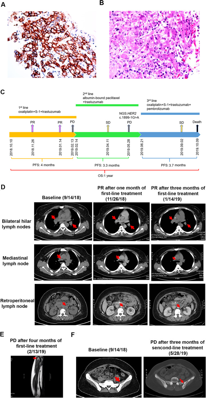
Figure 1.

A summary of patient's treatment history. (A): Immunohistochemistry staining analyses showed the tumor cells were positive for HER2 expression (3+). (B): H&E staining showed a poorly differentiated adenocarcinoma. (C): The entire treatment procedure. (D): Chest and abdominal computed tomography (CT) scans at baseline and in November 2018 and January 2019 demonstrating PR in bilateral hilar lymph nodes, mediastinal lymph node, and retroperitoneal lymph node. (E): Magnetic resonance imaging scans in February 2019 showed the patient developed metastasis to right humerus after failure of first‐line treatment. (F): CT scans in May 2019 showed the patient developed metastasis to pelvis after failure of second‐line treatment. Abbreviations: HER2: human epidermal growth factor receptor 2; NGS, next‐generation sequencing; OS, overall survival; PD, progressive disease; PFS, progression‐free survival; PR, partial response; SD, stable disease.
