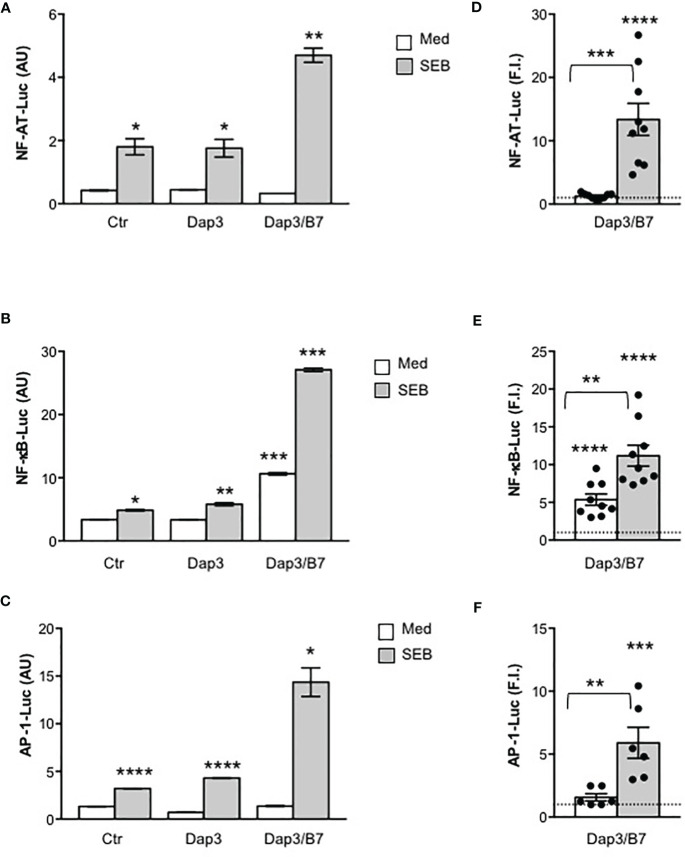
Figure 2.
Stimulation of CD28 by SEB induces transcription factor activation in the absence of MHC class II molecules. (A–F) CD28WT cells transfected with 5 µg GFP together with 10 µg NF-AT-luciferase (A, D), or 2 µg NF-κB-luciferase (B, E) or 10 µg AP-1-luciferase constructs (C, F) were unstimulated (Ctr) or stimulated for 6 hours with SEB (1 μg ml-1) alone or in the presence of Dap3 or Dap3/B7 cells. The results (A–C) are expressed as the mean of the luciferase units (AU) ± SEM after normalization to GFP expression and are representative of three independent experiments. (D) NF-AT luciferase activity (n = 9), (E) NF-κB luciferase activity (n = 9) and (F) AP-1-luciferase activity (n=8) of CD28WT cells stimulated with Dap3/B7 in the presence or absence of SEB. Bars show the mean fold induction (F.I) ± SEM after normalization to GFP values. Statistical significance was calculated by Student’s t test. Mean values: NF-AT, Dap3/B7 = 1.2, Dap3/B7 SEB = 13.3; NF-κB, Dap3/B7 = 5.3, Dap3/B7 SEB = 11.1; AP-1, Dap3/B7 = 1.5, Dap3/B7 SEB = 5.9. (*) p < 0.05, (**) p < 0.01, (***) p < 0.001, (****) p < 0.0001.

