Figure 7.
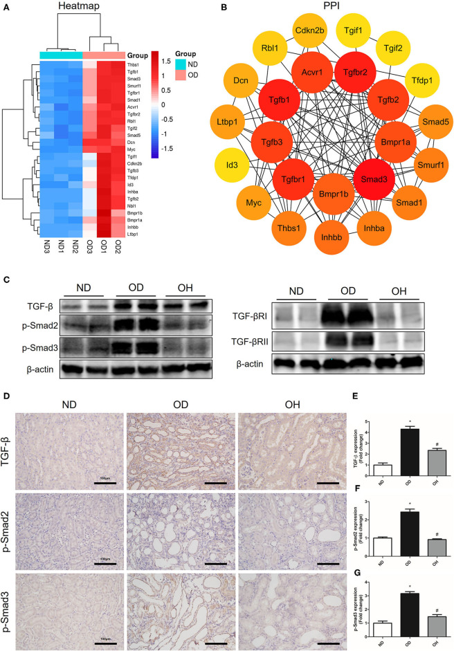
HRW administration suppresses oxalate-induced TGF-β signaling pathway activation in mice. (A) Heatmap and hierarchical clustering showing DEGs enriched in the TGF-β signaling pathway between the OD and ND groups. (B) PPI network constructed by DEGs enriched in the TGF-β signaling pathway. (C) The protein expressions of TGF-β, TGF-βRI, TGF-βRII, p-Smad2, and p-Smad3 in kidneys of mice were detected by western blotting. (D) The protein expressions of TGF-β, p-Smad2, and p-Smad3 in kidneys of mice were detected by immunohistochemistry (Scale bar = 100 μm). (E–G) Corresponding semiquantitative analysis of TGF-β, p-Smad2, and p-Smad3 immunohistochemistry staining. The results were expressed as mean ± SEM. Statistical comparisons were performed using a NewmanKeuls test (*p < 0.05 vs. ND group, #p < 0.05 vs. OD group).

