Figure 4.
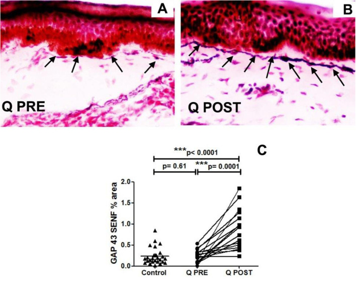
Immunohistochemistry results in skin biopsies for GAP3 nerve fibres. (A) NFCI skin biopsy section before treatment with Capsaicin 8% patch (Q PRE): sub-epidermal nerve fibres (arrows) were present, at a density similar to control/normal skin. (B) NFCI skin biopsy section in the same participant as above, post-treatment (Q POST); skin biopsy collected 3 months after Capsaicin 8% patch Qutenza (Q) application: note the marked increase of GAP 43 positive SENFs in length and thickness. Original magnification ×20. (C) Sub-epidermal GAP43 nerve fibres (SENFs; % Area) at pre-treatment visit (Q PRE) and visit 3 months after treatment (Q POST) with Capsaicin 8% patch Qutenza (Q). Statistically significant increase of SENFs at 3 months after Capsaicin 8% patch application (paired t-test). Statistically non-significant difference between control group and treated group before treatment (Q PRE), but statistically significant difference between control group and treated group after Capsaicin 8% patch application (Q POST) (Mann-Whitney test).

