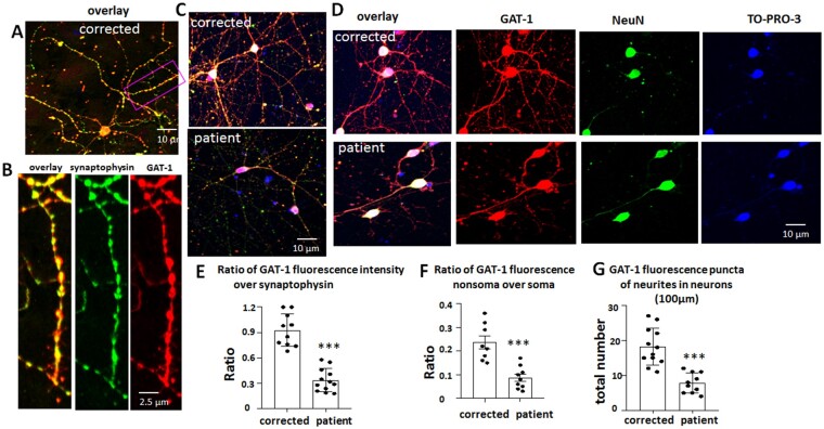
Figure 7.
Variant GAT-1(S295L) was retained in neuronal soma with reduced expression in dendrites. (A–D) GABAergic inhibitory neurons (aged 60–65 days) differentiated from CRISPR corrected and patient iPSCs were immunostained with rabbit polyclonal anti-GAT-1 antibody and mouse monoclonal anti-synaptophysin (A–C) or anti-NeuN antibody (D). In B, the boxed region from A is enlarged for better synaptic visualization. The rabbit IgG was visualized with Cy3 (red) and the mouse IgG was visualized with Alexa-488 (green). The cell nuclei were stained with TO-PRO-3 (1:500). (E) The raw fluorescence values of GAT-1 and synaptophysin in non-somatic region were measured and the ratio was plotted. (F) The raw GAT-1 fluorescence values in non-somatic region and somatic regions were measured and the ratio was plotted. The soma was identified by neuronal marker NeuN. (G) GAT-1 fluorescence puncta in neurons differentiated from the CRISPR corrected and the patient iPSCs were quantified. The total fluorescent puncta per 100 µm was measured. In E–G, n = 8–12 from four batches of differentiated cells. In E and F, average raw fluorescence of GAT-1 or synaptophysin from three non-selectively chosen areas in non-somatic region was measured for each neuron. The mean value was taken as n = 1. ***P < 0.001 versus corrected, one-way ANOVA and Newman-Keuls test. Values are expressed as mean ± SEM.

