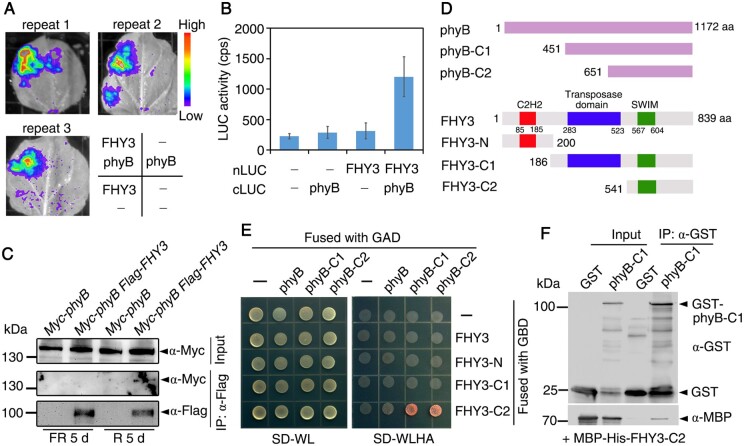
Figure 2.
phyB physically interacts with FHY3. A, LCI assay. FHY3 and phyB were fused with either the N- or C-terminus of luciferase (LUC). Different combinations of constructs were co-transformed into N. benthamiana leaves and LUC luminescence was monitored. B, Quantification of relative LUC levels (counts per second, cps) as shown in (A). Data are means ± SD, n=3. C, Co-IP assay. Seedlings were grown in red or far-red light for 5 d. Total proteins were immunoprecipitated with anti-Flag antibody followed by blotting with anti-Flag or anti-Myc. D, Diagram of FHY3 and phyB and their truncations. Numbers indicate positions of the amino acid residues. E, Yeast two-hybrid assay. FHY3 and its fragments were fused with the GAL4 DNA binding domain (GBD), whereas phyB and its fragments were fused with the GAL4 activation domain (GAD). SD-WL indicates synthetic dropout medium lacking Trp and Leu; SD-WLHA denotes synthetic dropout medium lacking Trp, Leu, His, and Ade. F, In vitro pull-down assay. After incubation, the recombinant proteins were immunoprecipitated with anti-GST antibody followed by blotting with anti-GST or anti-MBP.

