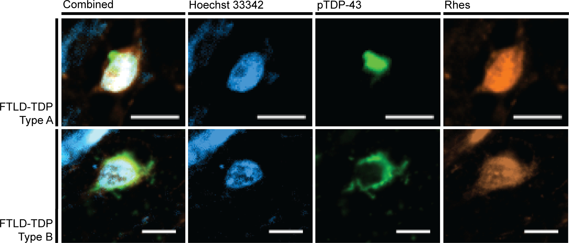
Figure 6:

Neurons positive for pTDP-43 (green) proteinopathy display a Diffuse Rhes (orange) phenotype. The FTLD-TDP Type A images come from the inferior frontal gyrus of a 66-year-old female with behavioral variant frontotemporal dementia. The FTLD-TDP Type B images come from the middle frontal gyrus of a 57-year-old male with behavioral variant frontotemporal dementia and amyotrophic lateral sclerosis. Scale bar represents 10 μm.
