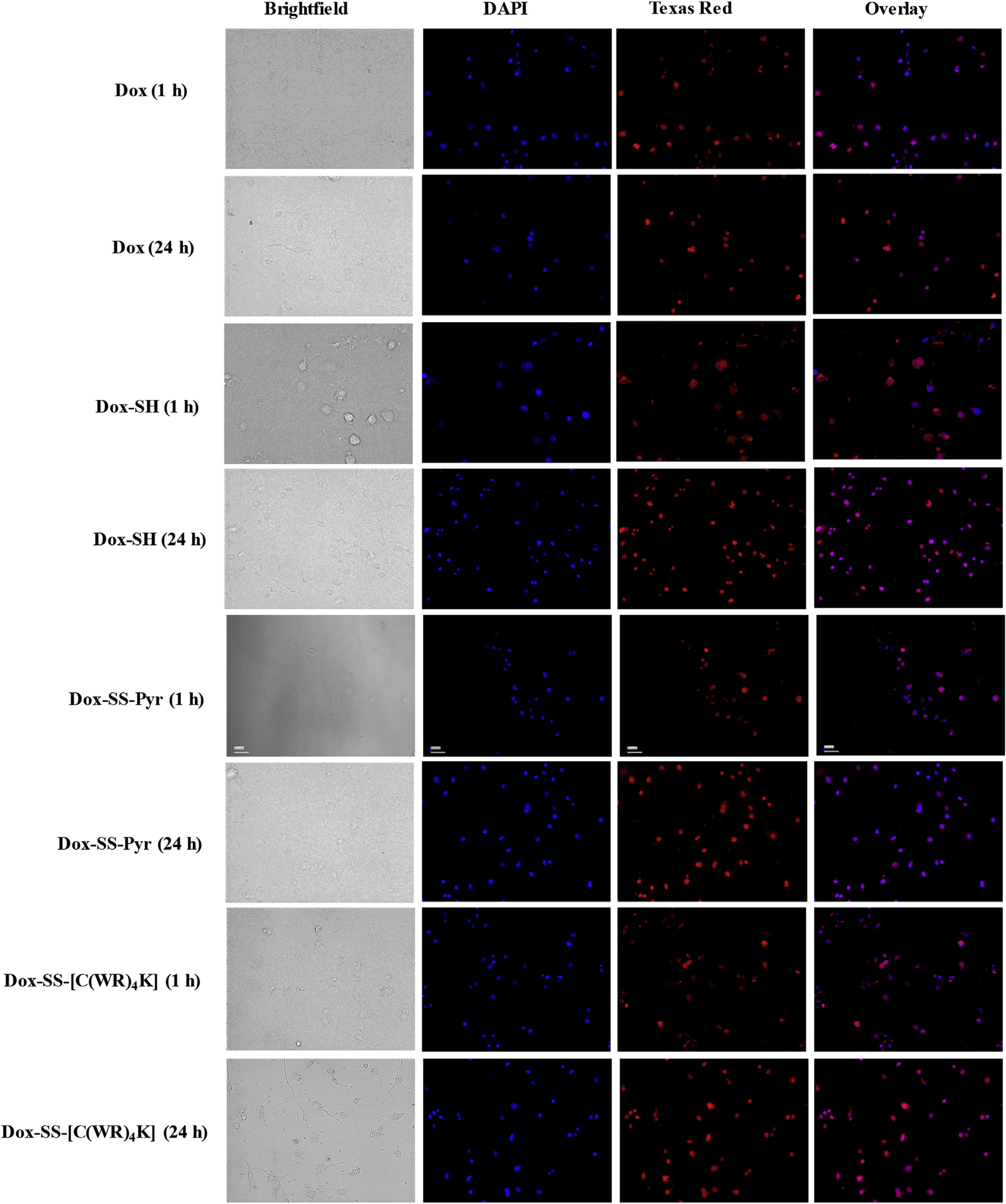
Fig. 5.

Fluorescence microscopy images of Dox (5 μM), Dox-SH (5 μM), Dox-SS-Pyr (5 μM), and Dox-SS-[C(WR)4K] (5 μM) uptake in MDA-MB-468 cells after 1 h and 24 h. Cells treated with the compound for 1 h or 24 h. Red represents the fluorescence of Dox. To identify the nucleus, the cells were dyed with DAPI.
