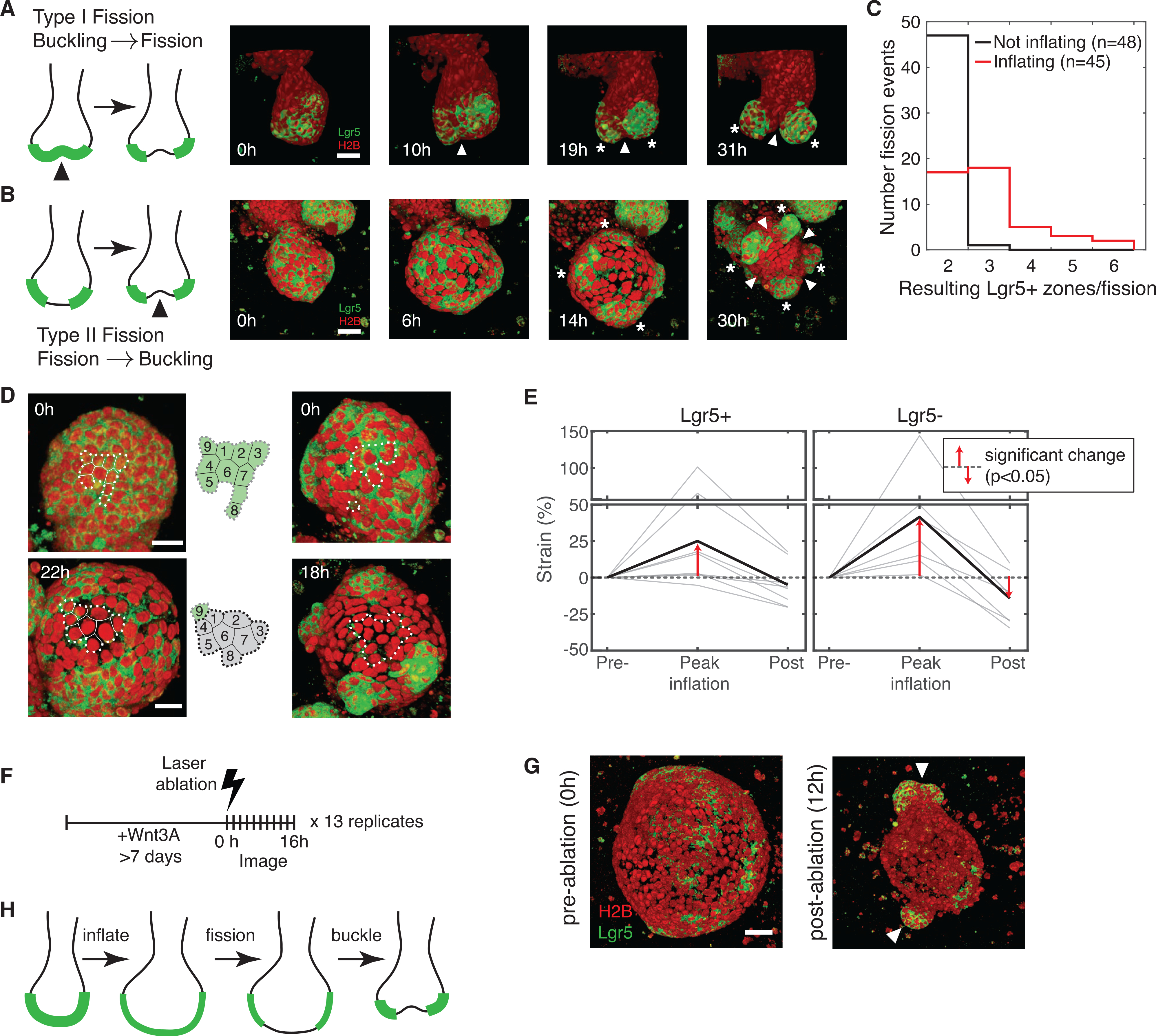
Figure 4. Stereotyped SCZ fission dynamics in intestinal organoids.

(A) Fission sequence (type I) by tissue buckling and invagination (white arrows) preceding Lgr5+ stem cell differentiation and fragmentation of SCZs (white asterisks). Red, H2B; green: Lgr5.
(B) Fission sequence (type II) with fragmentation of contiguous Lgr5+ SCZs preceding tissue buckling. The organoid bud appears inflated until 14 h. (A and B), arrows indicate invagination; asterisks indicate nascent SCZs. Scale bars, 20 μm.
(C) Number of nascent SCZs observed following fission from SCZs with inflated and non-inflated morphology.
(D) 40×-magnification and 6-min time lapse of organoid buds reveal that Lgr5+ cells differentiate in situ as organoid buds inflate. Scale bar, 40 μm. Dashed lines delineate a group of cells at early and late time points.
(E) The in-plane strain of the epithelium at three time points during luminal inflation and collapse. Gray, individual buds (n = 9); black, average. Strain is calculatedas the fractional change in mean inter-nuclear distance. Statistical significance by one-tailed t test.
(F and G) Laser ablation of cyst-like organoids grown in the presence of Wnt3A induces collapse, tissue buckling, bud formation, and localization of pre-existing Lgr5+ domains to nascent buds (white arrows). Scale bar, 50 μm.
(H) Summary of the model for type II SCZ fission.
