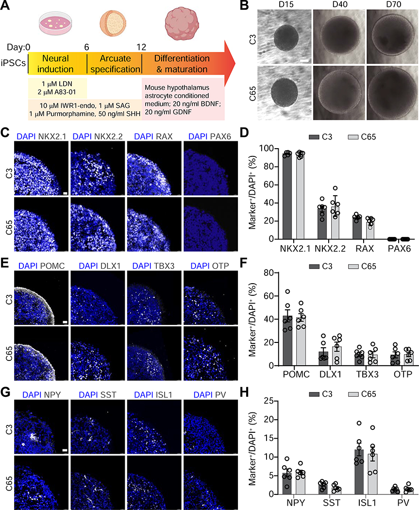
Figure 1. Generation and Characterization of Arcuate Organoids from Human iPSCs.
(A) Schematic describing the protocol for generating arcuate organoids (ARCOs) from human iPSCs.
(B) Sample bright-field images of ARCOs at 15, 40 and 70 days in vitro (DIV). Scale bar, 300 μm.
(C-H) Sample confocal images of immunostaining for NKX2.1, NKX2.2, RAX and PAX6 in ARCOs at 15 DIV (C, scale bar, 15 μm), for POMC, DLX1, TBX3 and OTP at 40 DIV (E, scale bar, 30 μm), for NPY, SST, ISL1 and PV at 40 DIV (G, scale bar, 30 μm), and quantifications (D, F, H). Values represent mean ± SEM with individual data points plotted (n = 6 organoids per iPSC line).

