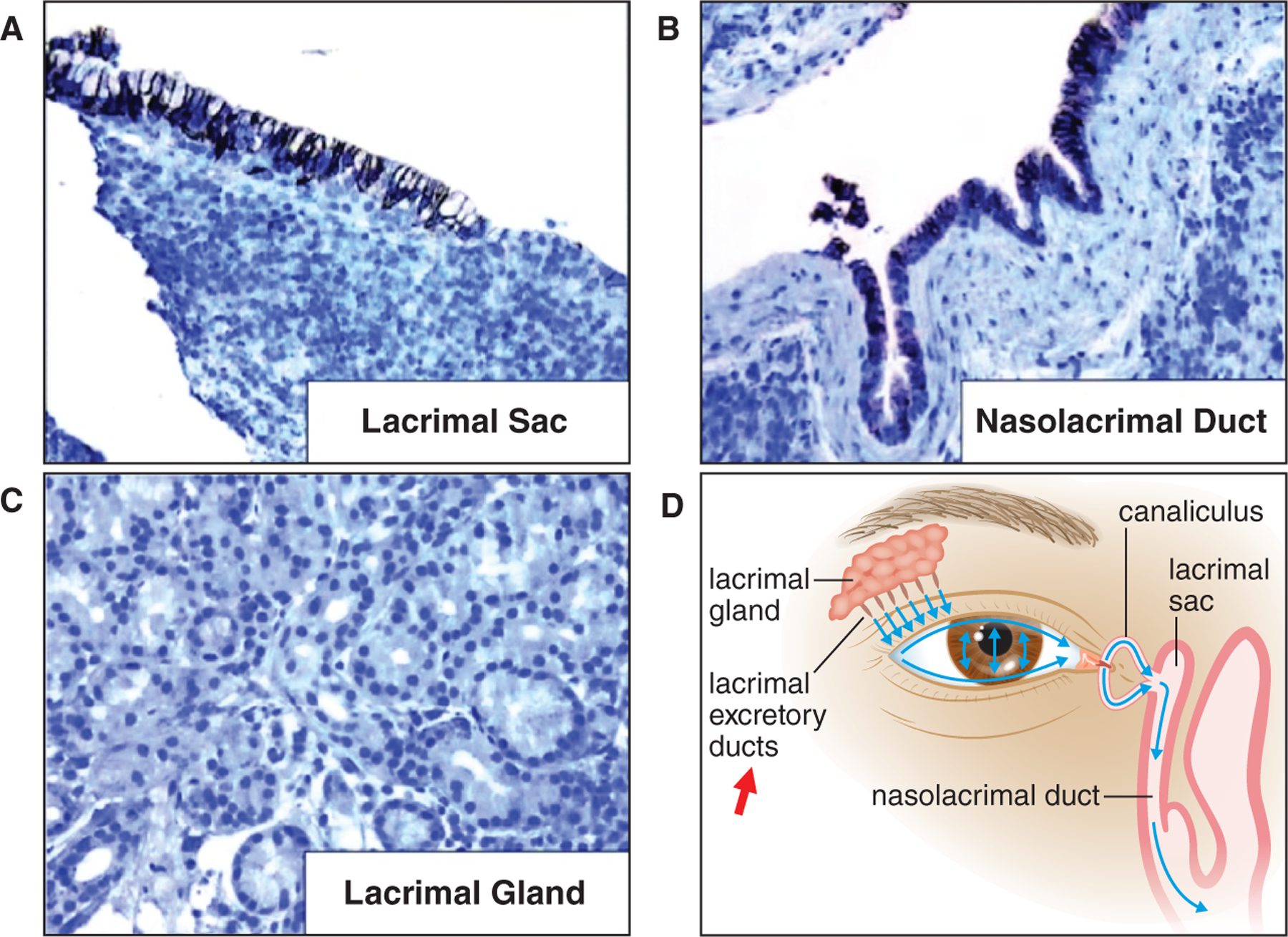
Figure 2:

NIS expression in normal lacrimal drainage tissues. NIS immunostaining (brown color) is detected at the basolateral membrane of the stratified columnar epithelial cells in (a) the lacrimal sac, and (b) the nasolacrimal duct. NIS immunostaining is absent in (c) lacrimal glands. NIS expression was not investigated in (d) lacrimal excretory ducts (red arrow).
