. 2021 May 21;19(9):1559–1570. doi: 10.1158/1541-7786.MCR-20-1017

Figure 5.

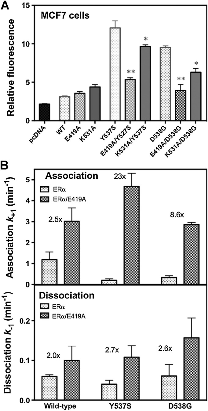
Figure 5. Ligand-binding kinetics and cellular activity of the ER. A, MCF-7 cells were transfected with plasmids for control, WT ERα and the eight indicated mutant ERs, as well as an ER-responsive luciferase plasmid, and constitutive transcriptional activity was monitored in the absence of added estrogen. The * indicates a significance of <0.05 and ** a significance <0.01. B, Ligand association and dissociation rates of the LBDs of WT, Y537S, and D538G ERα were monitored under pseudo first-order conditions using the fluorescent ligand, THC-ketone. Rate constants are shown in the presence of the Glu419–Lys531 salt bridge (gray bars) and in its absence due to the additional E419A (stippled bars). The fold increase in ligand association rate from removal of the salt bridge is indicated by the number above the bars for each receptor.

Ligand-binding kinetics and cellular activity of the ER. A, MCF-7 cells were transfected with plasmids for control, WT ERα and the eight indicated mutant ERs, as well as an ER-responsive luciferase plasmid, and constitutive transcriptional activity was monitored in the absence of added estrogen. The * indicates a significance of <0.05 and ** a significance <0.01. B, Ligand association and dissociation rates of the LBDs of WT, Y537S, and D538G ERα were monitored under pseudo first-order conditions using the fluorescent ligand, THC-ketone. Rate constants are shown in the presence of the Glu419–Lys531 salt bridge (gray bars) and in its absence due to the additional E419A (stippled bars). The fold increase in ligand association rate from removal of the salt bridge is indicated by the number above the bars for each receptor.